This is an Anatomy of the Ear template that can be used in a Biology lesson, as an example of a poster in the classroom, as a template for a study session, or as a visual for a textbook. The template is customizable with a few drags and drops in MyDraw.
Download Template:


Download Template:
What is the ear?
The ear acts as a receptor and filter in which auditory stimuli are transformed into information that is subsequently decoded by the brain. Also known as the vestibulocochlear organ its job is to detect, transmit and convert sounds into electric impulses. Or in other words, the main function of the ear is to maintain our sense of balance.
Structure and anatomy of the ear
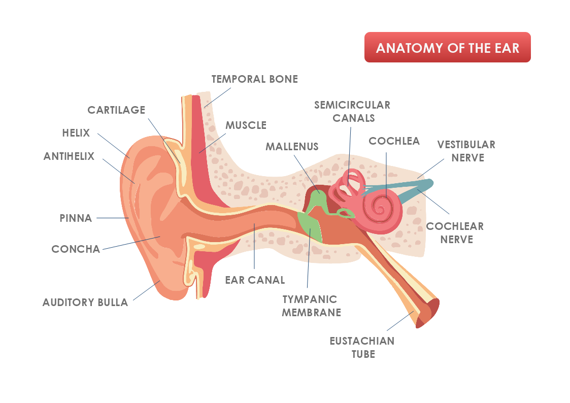
The external ear is structured by the pinna and the external auditory canal. Its function is to receive sounds and transmit them to the middle ear through the eardrum. The eardrum has a circular and flexible form, that starts to vibrate due to the received sound waves.
The middle ear is formed by the ossicles- the three minuscule bones named the malleus, incus, and stapes. The function of these bones is to form a bridge between the eardrum and the inner ear. The Eustachian tube connects the ear to the outer part of the nose and acts as an equalizing valve. This ensures that the pressure on either side of the eardrum is balanced and that sound can be heard correctly.
The inner ear includes the cochlea, a structure that has a spiral shape similar to a snail shell and is located in the bony labyrinth. The Organ of Corti transforms the mechanical energy of the sound waves into nerve energy by creating electric impulses that are sent to the brain through the auditory or vestibulocochlear nerve.
7 Interesting Facts about hearing
-
Without the pinna, there would be no way for sound waves to be funneled to the ear canal.
- The sound waves are the reason for our eardrum to vibrate.
- The movement of the 3 small bones in the ear is caused by vibrations of the ear drum.
- The hairs in the cochlear are tuned to respond to differences in sound frequency and pitch.
- The auditory nerve receives generated nerve impulses from the stimulation of the hair cells.
- The nerve impulses travel along the auditory nerve into the hearing center of the brain, known as the auditory cortex.
- The auditory cortex converts the nerve impulses into the sound that we hear.
How to create a custom shape in MyDraw?
- You can create your shape in a vector program and import/ insert it as a vector or raster image. The other option is to use MyDraw’s Basic shapes and connectors and make your custom shape.
- Once you are happy with the custom shape you could include it in your Library.
- Click on the textbook icon on the left side of the bar and choose “New Library”. Name the library and drag and drop the shape in it.
- Then right-click with the mouse on the Library tab and choose “Save as”.
- Choose the folder location on your computer and save it as a .nlb file.
- For future use, you could always add the newly created library to other diagrams.
- Once you have created your diagram/ template you can save the document in one of MyDraw’s native formats or export it in a preferred file format(PDF, SVG, EMF, VSDX, etc.).
- You can also export the document as a raster image.
