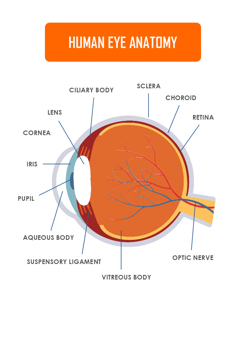
This is a Human Eye Anatomy template with labels that explains the structure of the human eye and is a great poster for an Anatomy class. The template is customizable with a few drags and drops in MyDraw.
Download Template:


Download Template:
Eye Outside the Eyeball
The Surface of the Eye- layers of the tear film keep the front of the eye lubricated.
The Front of the Eye- the muscles of the iris, widen or narrow the pupil to control the amount of light that reaches the back of the eye.
The Back of the Eye- the photoreceptors change the light into energy that is transmitted to the brain.
Did you know that?
Humans have two eye visions and not one.
Humans have a horizontal field of view of about 1500 with one eye and about 1800 with both eyes.
Our eyes are like cameras that capture colorful images in seconds, which are processed by our brains as messages.
Each eye can see slightly different images.
If you close one eye and the world looks flat-two-dimensional.
How to create a custom shape for the Human Eye Anatomy diagram?
- You can create your shape in a vector program and import/ insert it as a vector or raster image. The other option is to use MyDraw’s Basic shapes and connectors and make your custom shape.
- Once you are happy with the custom shape you could include it in your Library.
- Click on the textbook icon on the left side of the bar and choose “New Library”. Name the library and drag and drop the shape in it.
- Then right-click with the mouse on the Library tab and choose “Save as”.
- Choose the folder location on your computer and save it as a .nlb file.
- For future use, you could always add the newly created library to other diagrams.
- Once you have created your diagram/ template you can save the document in one of MyDraw’s native formats or export it in a preferred file format(PDF, SVG, EMF, VSDX, etc.).
- You can also export the document as a raster image.
