

The heart is divided into four chambers and it is one of the most important organs in the human body. The heart is like a network of blood vessels that pumps blood throughout your body. Its function depends on the family history, personal health history, and lifestyle all affect how well your heart works.
7 Facts about the Function and Anatomy of the Heart
The heart is a muscular organ that is responsible for pumping blood throughout the body. It is located in the chest cavity, behind the sternum, and between the lungs.
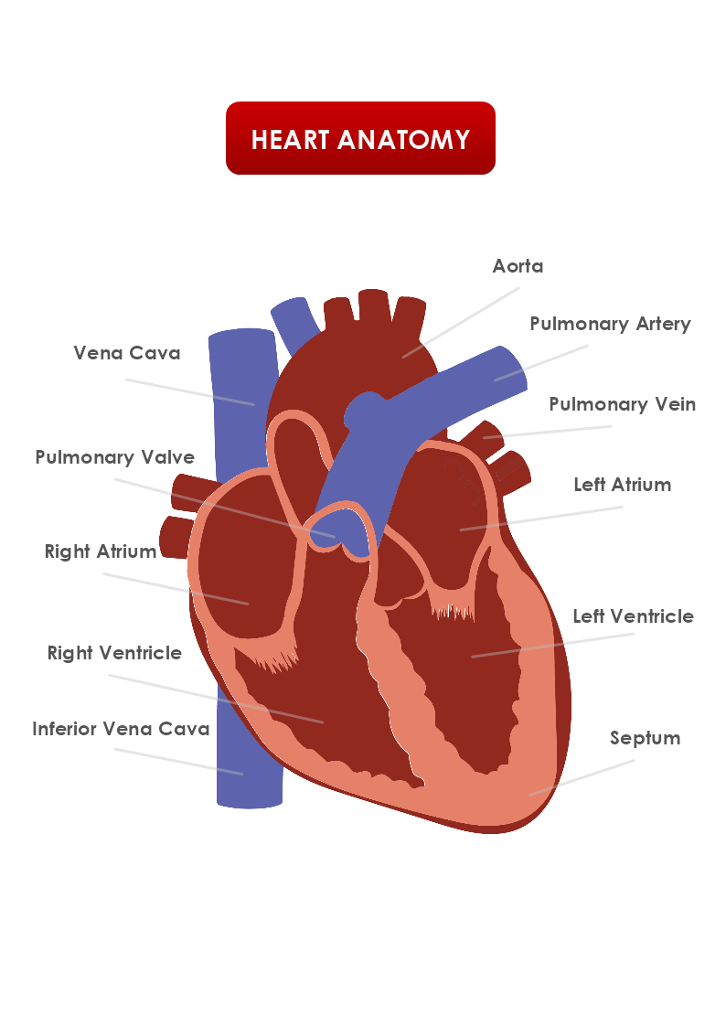
It has four chambers: the left and right atria, and the left and right ventricles. Oxygen-poor blood from the body enters the right atrium and is then pumped into the right ventricle and sent to the lungs for oxygenation. Oxygen-rich blood from the lungs returns to the left atrium and is then pumped into the left ventricle and sent out to the rest of the body.
The heart has its electrical conduction system, which helps to coordinate the contraction of the heart muscle and ensure that blood is pumped efficiently. The sinoatrial (SA) node, located in the right atrium, acts as the heart's natural pacemaker and sends out electrical signals to stimulate contraction.
The heart has a network of blood vessels, including the coronary arteries, which supply the heart muscle with oxygen and nutrients. If these arteries become blocked or narrowed, it can lead to a condition called coronary artery disease, which can cause a heart attack.
The heart has three layers of tissue: the outer epicardium, the middle myocardium, and the inner endocardium. The myocardium, or heart muscle, is responsible for pumping blood and is made up of special cardiac muscle cells.
Four valves help to ensure that blood flows in the correct direction through the heart. The tricuspid valve is located between the right atrium and right ventricle, the pulmonary valve is located between the right ventricle and the pulmonary artery, the mitral valve is located between the left atrium and the left ventricle, and the aortic valve is located between the left ventricle and the aorta.
It is a vital organ that is essential for maintaining the circulation of blood and oxygen to the body's tissues. It can adapt to changes in the body's needs, such as during exercise, and can increase or decrease its rate of contraction to meet these needs.
